Diagnosis: Right bundle branch block (RBBB) with Left anterior fasicular block (LAFB)
Discussion: Amongst the criteria for RBBB is an RS Rprime in V1, and wide S waves in leads I and V6. The RS Rprime can be broken down as follows; the R wave is caused by the normal septal depolarization which is directed towards V1, since it occurrs from left to right across the septal wall. The S wave is caused by LV depolarization which is directed away from V1. The Rprime is caused by the late depolarization of the RV, which is again directed towards V1. Normally the RV depolarizes much earlier and contributes to the initial R wave along w/ the septal depolarization..
Another consequence of late RV depolarization is the wide S waves in leads V6 and I. The force vector from RV depolarization is directed anteriorly and to the right. This vector then will register as a negative deflection in V6 and lead I. Since this RV depolarization is now occurring late it adds to the S wave in I and V6. Also, since the RV is being depolarized by current that must traverse normal muscle before getting to the right sided conduction system, this depolarization occurrs slower and thus also contributes to the widending of the S waves in V6 and I. It also tends to prolong the QRS duration, which is also seen in this case.
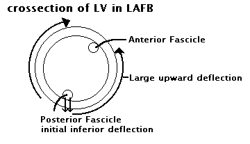
In LAFB characteristically one sees an extremely left sided axis. (note there are no discrete anterior and posterior fasicles in humans, but much of the original work was done in dogs who have discrete fasicles). In LAFB the posterior wall of the LV wall must depolarize first through the posterior fasicle and then current courses from posterior to anterior to depolarize the rest of the myocardium of the LV. This causes an initial small inferior current vector then a much larger upward current vector. In inferior leads then we see a small R wave followed by a large S wave. Also in leads I and AVL we see a small Q wave followed by a large S wave. If you see extreme left axis i.e. -60 or more and no LVH or other reasons for LAD, then it is most probably LAFB.
The diagram below shows the movement of the force vectors in the left ventricle during depolarization in a pt w/ LAFB.
Hit the Menu button at the top of the screen to return to the menu page.
note: The explanations contained here are based upon my own understanding of Electrophysiology as a 4th year medical student. I am pretty sure I am right, but if you have more to add, comments, questions or corrections please email me.