

NetMedicine Radiology Library- version 0.2
By:
Ashraf H. Nashed, M.D.- Assistant Director, Residency in Emergency
Medicine, Morristown Memorial Hospital
Ravi Murthy, M.D.- Department of Radiology, Morristown Memorial
Hospital
Manuel Correia, Jr- University of Medicine and Dentistry of New
Jersey, New Jersey Medical School
Glenn Fink, M.D.- Director of Medical Informatics, Department
of Emergency Medicine, Saint Barnabas Medical Center
NetMedicine has created an extensive Radiology Teaching Library on the
Web containing hundreds of images. Save the images on your computer
for future reference or for teaching purposes!
Hints for better results in viewing these images.
Select from the following or scroll below to find the images you
are interested in.
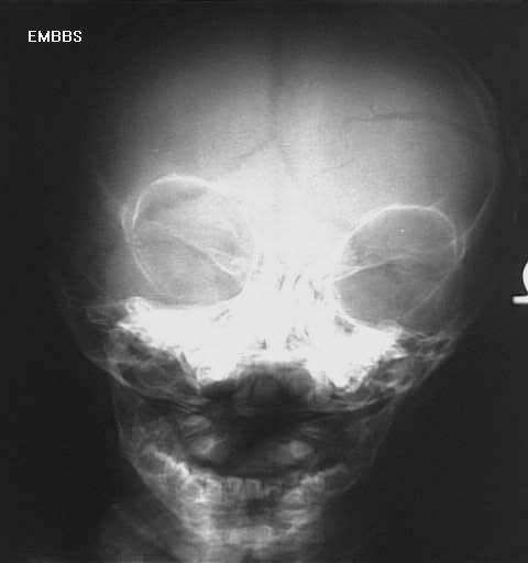
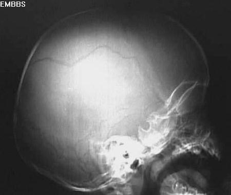
Head
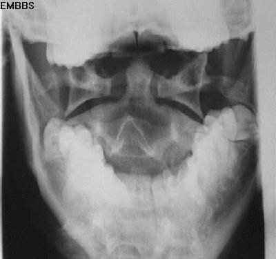
Skull
Facial Bones
Mandible
Nasal Bones
Neck
Soft tissue
Cervical Spine (see spine)
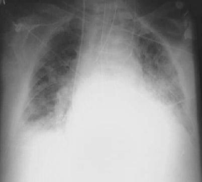
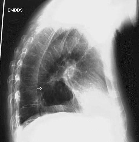
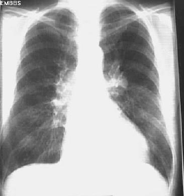
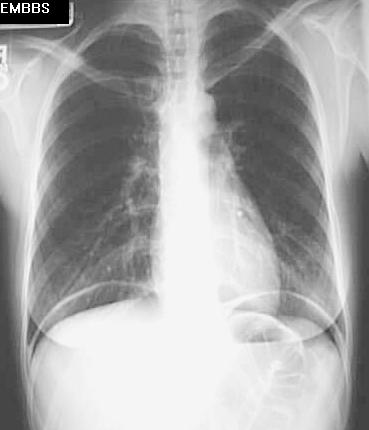
Chest
Thoracic Spine (see spine)
Spine
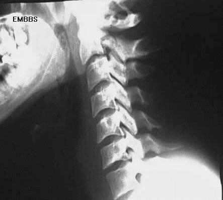
Cervical
Thoracic-under construction
Lumbosacral-under construction
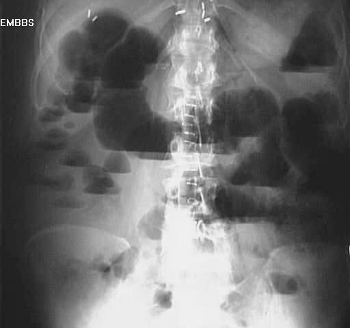
Abdomen
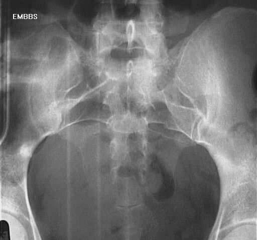
Pelvis
Upper Extremities
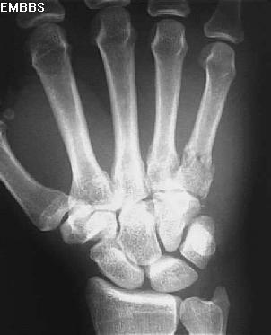
Hand
Wrist
Forearm
Elbow
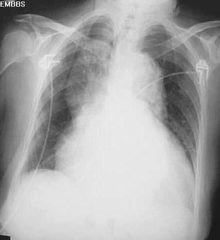
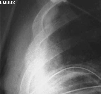
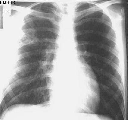
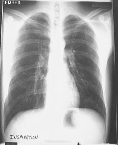
Shoulder
Scapular fracture
demonstrated on the lateral border.
Humeral surgical neck fracture, displaced.
.
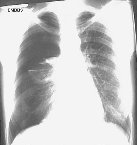
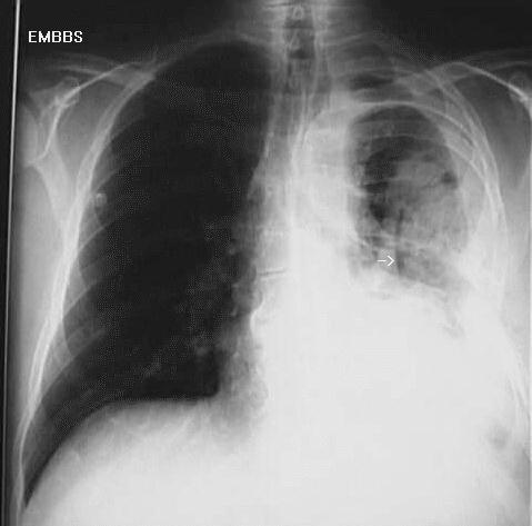
Posterior shoulder dislocation, axillary view, note the humeral head is not in the glenoid fossa, AP with internal rotation, note the small and false "joint space" as the humeral head sits behind the glenoid fossa, external rotation, Y view
.
Lower Extremities
Hip
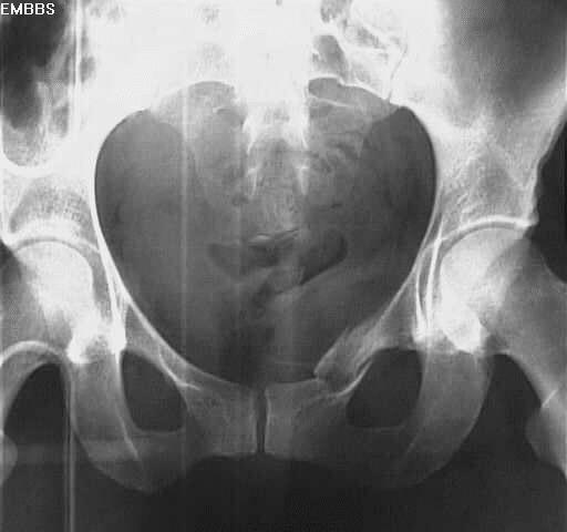
Intertrochanteric fracture-AP
, close up
, Lateral
Hip frx, infant with bone scan
Femur
Femoral shaft fracture
Knee
Patellar frx with disruption of the patellar ligament
.
Tibial plateau fracture AP, oblique
.
Tib/Fib
Massoneuve's fracture- Lateral view
, Oblique view
, full AP view
, full Oblique view
Massoneuve's fracture- posterior tibial frx-Lateral view
, AP view with widening of the syndesmosis
, both frxs
, close up of prox fib frx
, close up of distal tibial frx
, AP view of the distal leg
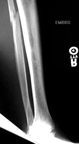
Tib/Fib fracture-AP
, Lateral view
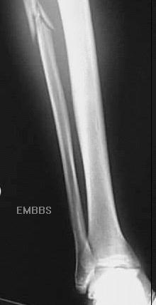
Tib/Fib fracture-AP distal
, AP prox
imal
Fibular fracture AP
, Lateral
Tibial frx - displaced Salter II
, AP
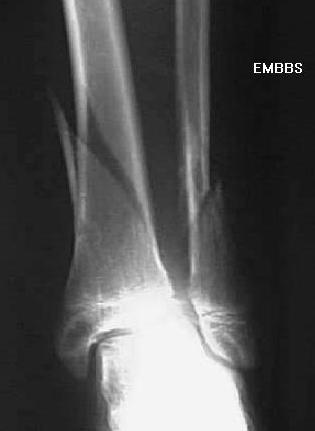
Ankle
Bimalleolar fracture
Trimalleolar fracture-ap
, Lateral view
Ankle dislocation with distal fib frx, medial malleolar frx
Foot
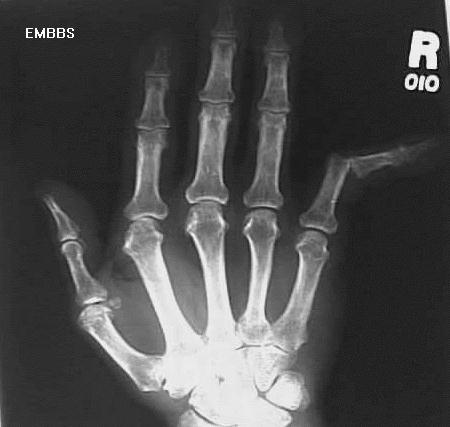
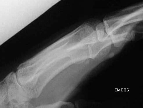
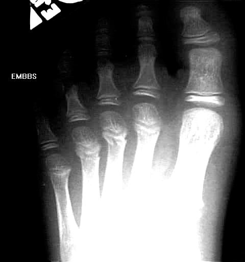
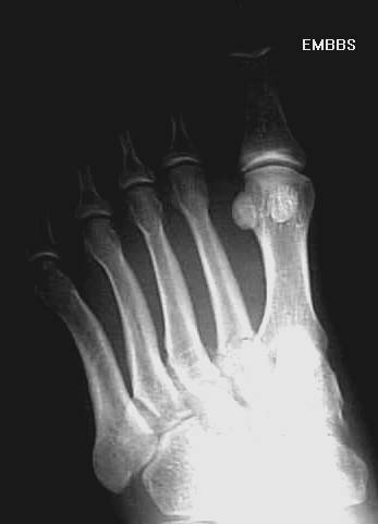
Metatarsal fractures, of the distal 2nd, 3rd, 4th metatarsals (AP view)
, Oblique view
Dancer's fracture, AP view
, Oblique view
, Lateral view
Lisfranc frx-Lateral view
, Oblique view
, AP view
, close up
Metatarsal dislocation, first metatarsal-Lateral view
, close up
, AP view
, close up AP
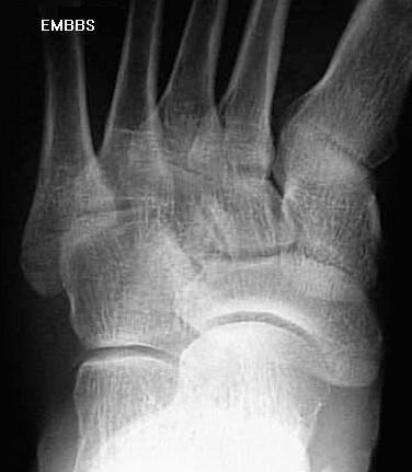
Fracture of cuboid-Oblique view
, close up of oblique view
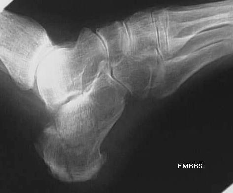
Fracture of the calcaneus-Lateral view
, post op fixation- Lateral view
Metatarsal fracture, base of the 5th metatarsal- Oblique
, close up
Calcaneus fracture - axial view
, Lateral view
Metatarsal stress fracture- 4th metatarsal
, close up
Calcaneal osteomyelitis
, Close up
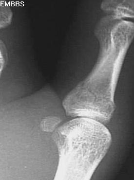
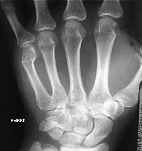
Fracture base of second metatarsal, AP view, close up view.

NetMedicine Home Page
Downloading and using one of these free programs as a helper application will significantly improve JPEG image quality.
Copyright ©1996, Triple Star Systems,
Inc. All Rights Reserved.
Last updated by Glenn Fink, M.D. 9/6/96
)window.location='http://www.netmedicine.com/xray/img_xr/foot4az.jpg')