Skin examination
Colour
Moisture
Temperature
Efflorescence
Trophics
Turgor
Oedemas
Skin adnexa
The skin is rosy, warm, and elastic, having no continuity defects.
When examining the skin by inspection and palpation we concentrate
on colour, moisture, temperature, turgor, presence of pathological efflorescence,
bleeding manifestations, and oedemas.
Colour
Moisture
Enhanced moisture depends on enhanced perspiration.

Localised moisture
in armpits, on palms, and soles, occurs in people with neurovegetative
dysbalance, commonly accompanied by acrocyanosis and acrohypothermia.
Diffuse moisture
on the whole body surface is present in lytic temperature decrease,
thyrotoxicosis, shock, and hypoglycaemia. Nocturnal sweating can be
related to malignant tumours and tuberculosis.
Reduced moisture
Localised form occurs in ischaemia.
Diffusion form can
be found in dehydration and cachexia. The skin is dry and wrinkled
and peels off.
Temperature
Body temperature depends on the blood supply of the skin, it can
be tentatively assessed by touch of hand.
Locally decreased temperature
is characterised by pallid; cold skin (could be cyanotic) as a result
of impaired blood supply (ischaemic disease of blood vessels of lower
extremities, Raynaud's disease).
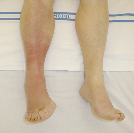
Locally increased temperature
is characterised by reddening and oedema of the skin and is caused by
inflammation (erysipelas, thrombophlebitis).


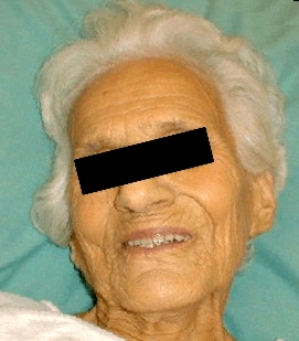
Anaemic face
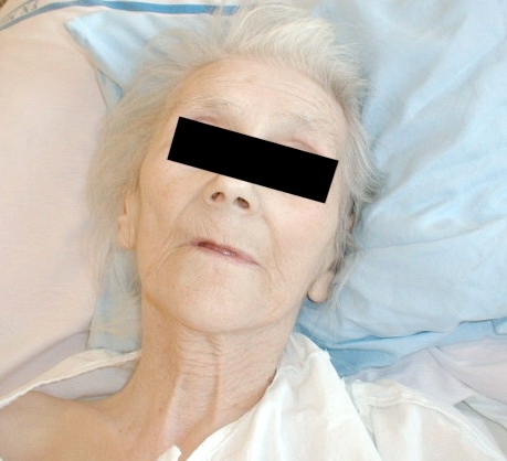
Pallid skin
Facies mitralis
Erysipelas

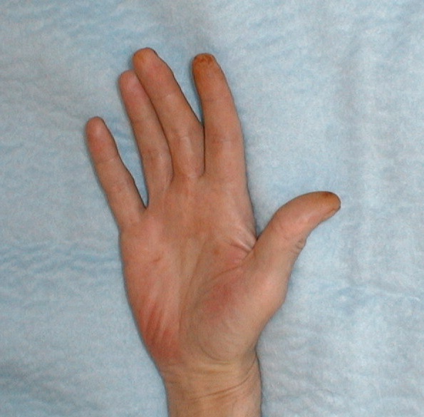
Dupuytren's
contracture
palm erythema

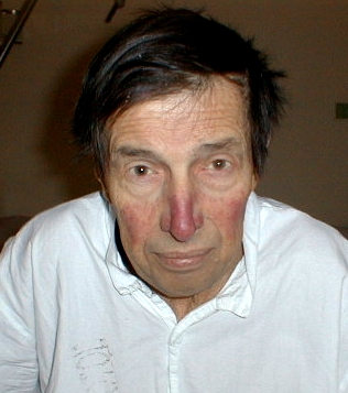
Face cyanosis -
congenital
heart disorder
Face skin
icterus
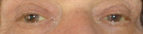
Sclera and face
skin icterus

Icterus
Icterus
Icterus
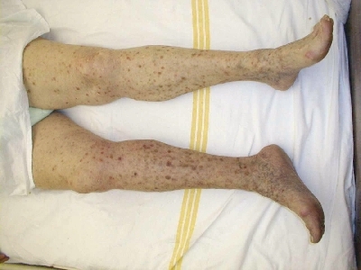
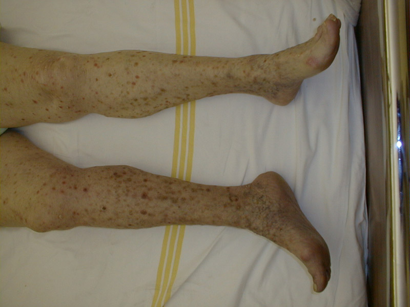
Shank
hyperpigmentation
Skin efflorescence
cannot be found on the skin of a healthy person. Its presence
is the sign of a skin disease or can be the secondary manifestation
of the infectious or internal disease. Dermatological
terminology is used for describing.
macula = area blot
papule = protruding blot
vesicula = blister filled by clear liquid
pustule = blister with turbid liquid
Findings can transform continuously. Exact description, localisation, and configuration, and even the dynamics of the disease are required for judgement.
Some diseases are accompanied by distinctive findings:
Scarlet fever (scarlatina): small-macular red
exanthema is localised on the skin of the abdomen, it spreads
onto the legs and the rest of the body; it does not appear
at the vicinity of the mouth.
If untreated, the disease can lead to skin exfoliations.
Measles (morbilli): macular exanthemas localised
initially on the face and neck; they tend to merge together later.
There are so called Koplik's spots
at the mucous membrane of the mouth.
Chickenpox (varicella): begins as a macular,
later vesicular exanthema on the surface of the whole body (including
areas with hair), gradually it dry out. Eruption of efflorescence runs in
the cycles.
Shingles (herpes zoster): vesicular, later pustular efflorescences
are arranged in the groups that follow peripheral nerves route, but
also branch of the nervus trigeminus. The disease is caused by
the varicella - zoster virus in adult patients weakened by other diseases
(e.g. tumours).
Cold sore (herpes labialis, nasalis): vesicular
or pustular efflorescences are found on the lips, below the nose
or by the nose orifices in febrile diseases (croupous pneumonia, viral
infections), or in insolation.
Allergic exanthemas take the form of either urticarial
(nettle-rash) exanthema or their appearance may resemble findings present
in infectious diseases. In that case, they are called according to the disease
they resemble (e.g. morbiliform, scarlatiniform etc.) Itchy white or rosy
buds of a map-like appearance are typical for urticaria. Allergic exanthemas
manifest as local affections, most commonly caused by direct contact (plants,
cosmetics), or generalised affections of various appearance - on the skin
of the trunk and limbs. They tend to mingle and their eruption is recurrent
(drugs, food).
Transient oedematous swelling on the face, neck, or perhaps other areas is the sign of Quincke's oedema.
Erythema nodosum are specific painful red and
violet infiltrates located on the shanks (sarcoidosis, idiopathic intestinal
inflammations, or the origin may be unclear).
"Butterfly exanthema" is distinguished by symmetrical reddening of the face that is distinctively shaped (lupus erythematosus).
Osler nodes are bright, red coloured lentil size
nodes, which can be found on the fingertips. They are caused by mycotic
micro-embolisation in infectious endocarditis.
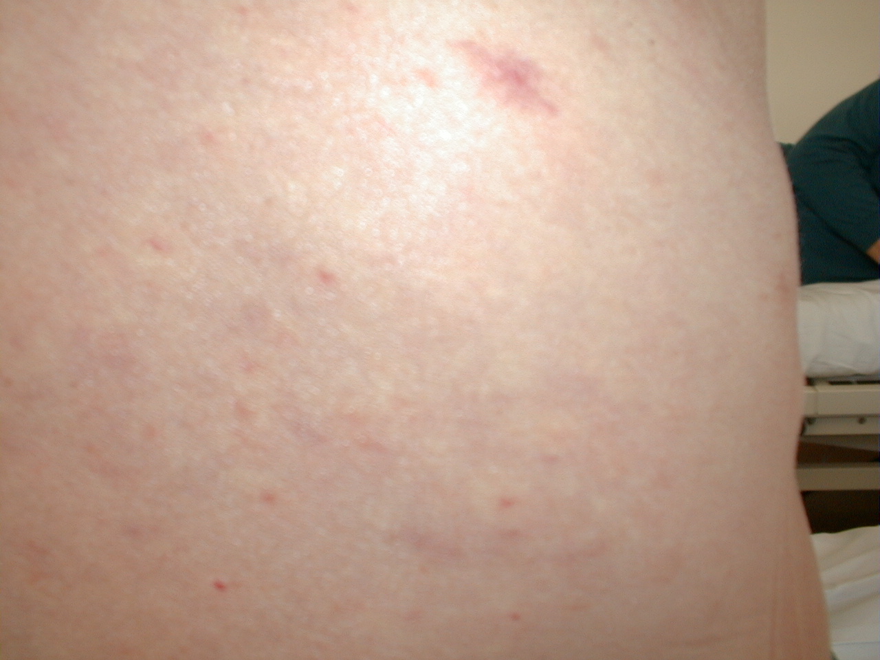
Various morphological findings in the form of petechiae, haematomas,
maculopapular efflorescences, or area infiltrations can all represent evolutionary
changes of vasculitis.
Xanthelasma is a shallow protruding area on the eyelid, close to the nose. It is caused by the accumulation of fat (hyperlipoproteinaemia, rarely in a healthy person too).
Xanthoma (tuberosum) is generally larger, commonly located on the muscle
tendons (some hyperlipoproteinaemias).
"Naevus arachnoideus" (spider angioma) is red,
made of a central arteriole wrapped by venules into periphery. Usually,
they are located in the upper part of the trunk and in the face.
In more advanced cases of hepatic cirrhosis they can appear on the arms
as well (they may appear non-specifically e.g. during pregnancy). When subjected
to pressure they become anaemic.
Haemangiomata are most commonly of lentil appearance,
but also they may be of irregular shape, at various locations in elderly
people.
Bleeding manifestations (haemorrhagic diatheses) on the skin
and mucous membranes arise spontaneously in cases of primary and secondary
haemocoagulation disorders.
Petechiae are ecchymoses, dotty haemorrhages in thrombocytopenia, thrombocytopathia, and vasculitis.
Purpura arises of multiplex petechiae.
Haematoma has its origin in substantial subcutaneous
bleeding in case of e.g. coagulopathy. They gradually decolourise over time
(haemophilia, incorrect anticoagulation therapy, blunt trauma, hepatic cirrhosis).
Maculo-papular
skin changes
Papule

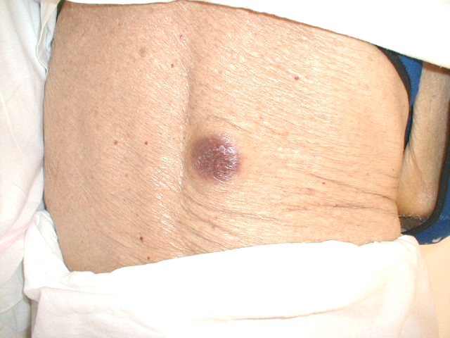
Allergic skin
reaction cased
by plaster
![]()
Allergic skin
reaction cased
by plaster
(on the hip)
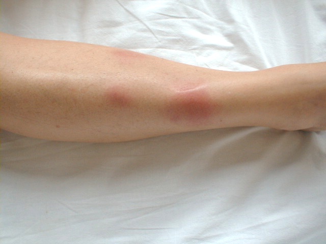
Erythema nodosum
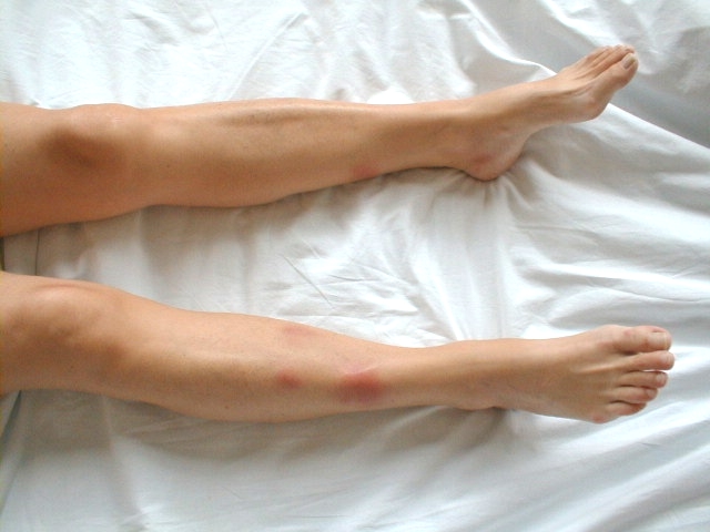
Erythema nodosum
on the legs
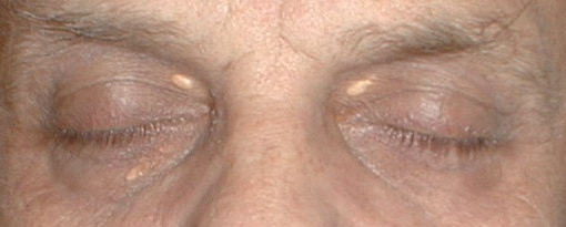
Xanthelasmata

Spider angiomata
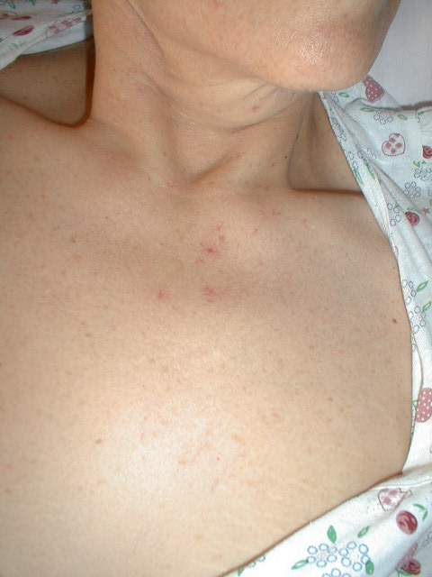
Spider angiomata
![]()
Spider angiomata
Detail of a
spider angioma
Vasculopatia
![]()
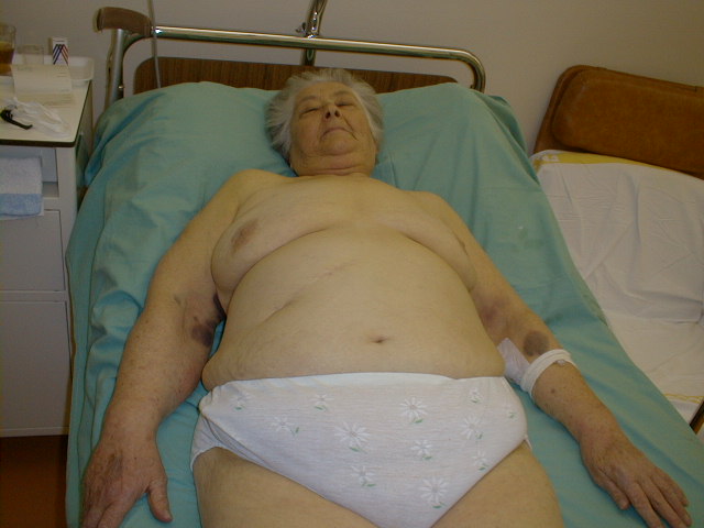
Large haematomas
on the chest and
arms
Haematomas
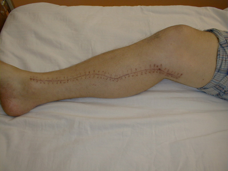
Postoperative
scars have distinctive shapes and localisations. The appearance
and colour allow to estimate the type of operation, history of healing,
and the time elapsed since opening the skin.
So called keloid scars are bulging, protruding,
reddish, found in person with individual redisposition.
Post-injury scars are irregular, in various
locations.
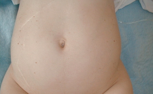
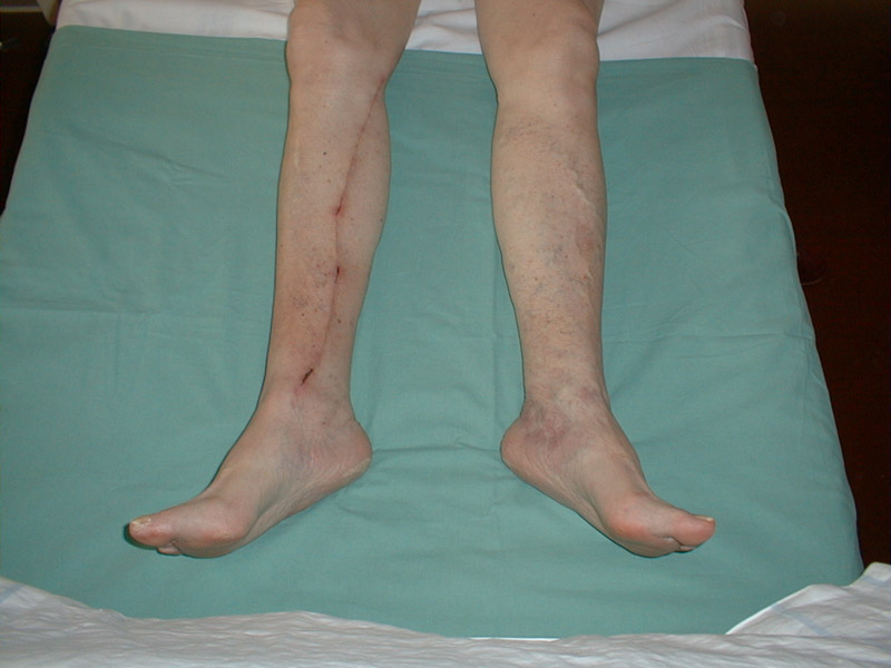
Scars
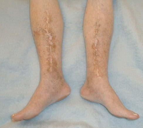
Scars on the
legs caused by
extraction of
venous grafts
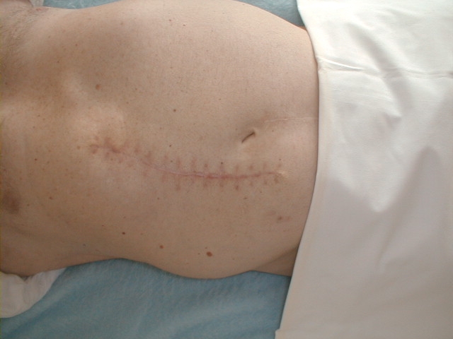
Scar after surgical
revascularisation
of myocardium
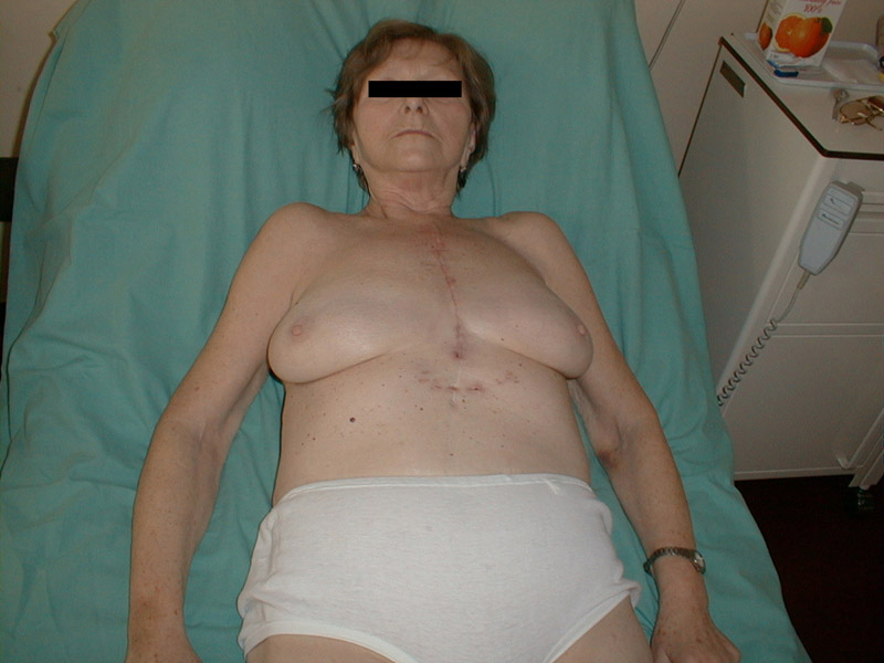
Scar after
sternototomy
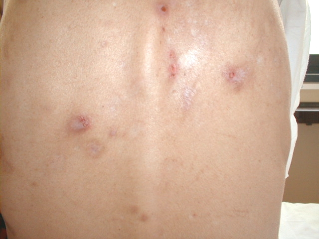
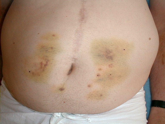
Scars of healed
furuncles
(back of a woman
suffering from
diabetes)
Trophic skin changes
are caused by vascular (ischaemic) and innervation disorders.
| |
Bedsores (decubitus) are the most common. They constitute in immobile patients on the heels, and sacral and gluteal areas first as a superficial local ischaemia, gradually worsening to necrosis. |
| |
Varicose ulcers localised on shanks are of various shapes, sizes, and depths and can be observed in patients with chronic venous insufficiency. |
| |
In chronic ischaemia trophic skin defects on the toes (ischaemic disease of blood vessels of lower extremities, diabetic microangiopathy) can be observed. |
Skin turgor
depends on hydration of the skin, the epidermis and its structure.
Decreased turgor is common in older age and is caused by decreased elasticity of epidermis.
In other cases dehydration caused by fluid loss contributes to decreased turgor (decompensated diabetes mellitus, diabetes insipidus, intensive diuretic therapy) or dehydration can be caused by insufficient intake of fluids (reduced thirst feelings in elderly people). The combinations of both causes are frequent, too.
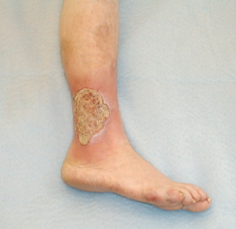
Shank ulcer
Large
shank ulcer
Trofic defect
on the big toe
of a diabetic
![]()

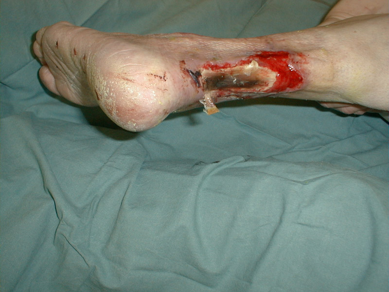
State after the
amputation of
the toes in a
diabetic
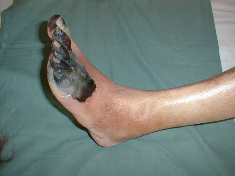
Diabetic foot

Gangrene of the
right leg, detail
![]()
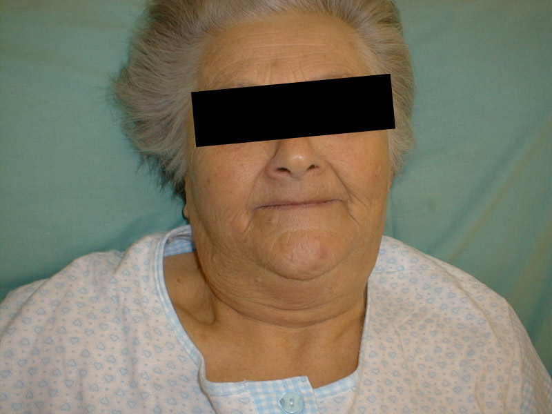
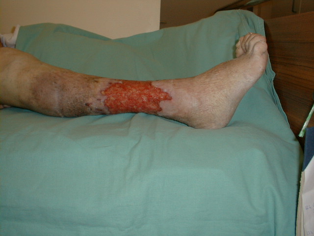
Secondary
skin changes
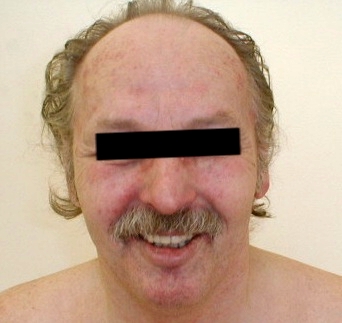
Secondary
skin changes
on the face
Oedemas
Oedemas are caused by an accumulation of extracellular fluid in the interstitium.
Local or generalised oedemas can be recognised.
Local oedemas
Inflammatory oedemas appear in the site of inflammation. The oedema is painful; the skin is warm and erythematous.
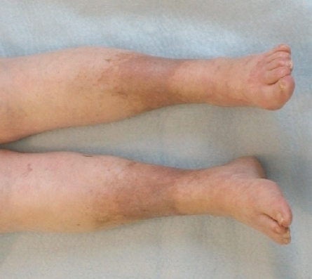
Venostasic oedemas occur in the blockage
of the venous system (phlebothrombosis).
The skin is taut, sensitive, palpation causes a shallow
dimple; cyanosis can be observed.
Lymphoedemas are caused by
the obstruction of lymaticph vessels or nodes by tumours, metastases,
or parasites.
The skin is pallid, rigid, and painless. After palpation, no
dimple occurs. The long-lasting obstruction causes induration
of the epidermis.
Allergic oedemas
can be found anywhere in the body, including mucous membranes
(Quincke's angioneurotic oedema, contact allergy, insect stings).
They tend to be flat, painless; they keep the colour and temperature
of the surrounding skin. Even eyelid oedemas in patients with
acute glomerulonephritis are considered of allergic origin.
Systemic oedemas occur in case of massive fluid
retention. From etiopathogenetic point of view there is various participation
of venostatic constituent, hypoproteinaemia and changes of vessel wall permeability.
Cardiac oedema occurs in
case of the right heart insufficiency. In walking patients they
constitute in area perimaleolaris; they advance to the shanks
and thighs. In recumbent patients they are found on the shanks,
the lower part of the thighs and in the loins. In the most
severe cases they stretch to the abdominal area and they affect
the outer genitals. Ascites, hydrothorax, or hydropericarditis
occur. The state is called anasarca.
Renal oedemas can be found
in nephrotic syndrome. They occur on the eyelids,
in the face, on the genitals, and in lumbosacral parts of
back.
Hepatic oedemas manifest
in decompensated hepatic cirrhosis. Ascites is predominant, but lower
extremities oedemas may occur as well.
Hypoproteinaemic oedemas in case of
hypalbuminaemia are soft, with persisting dimple after palpation.
Myxoedemas
form by accumulation of mucopolysacharides in the face and forearm
("iron sheet forearm"); they are of tough consistence.
Hair
has typical appearance and position depending on the sex.
Thin hair can be found in both sexes in hypogonadism,
hypopituitarism, hypothyroidism, and hepatic cirrhosis
and in males treated by oestrogens.
Stronger and denser hair (hypertrichosis,
hirsutism) is important in women. Mild forms
can be observed in older women on the face and in case of Cushing's
syndrome. More severe forms accompany androgenic tumours of the adrenal
cortex and androgen treatment (doping!).
Alopecia
is diffuse or local loss of hair. It occurs in cytostatic treatment,
in abdominal typhus, and thyrotoxicosis. In some men, the diffuse
alopecia is a common finding. Local alopecia (alopecia areata)
is rather rare to find.
![]()
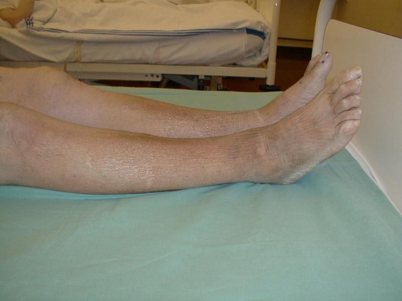
Venostatic oedemas,
ileofemoral
phlebothrombosis
of the right leg
![]()
![]()
Lymphatic oedemas
![]()
Lymphoedema of the
lower extremity,
detail
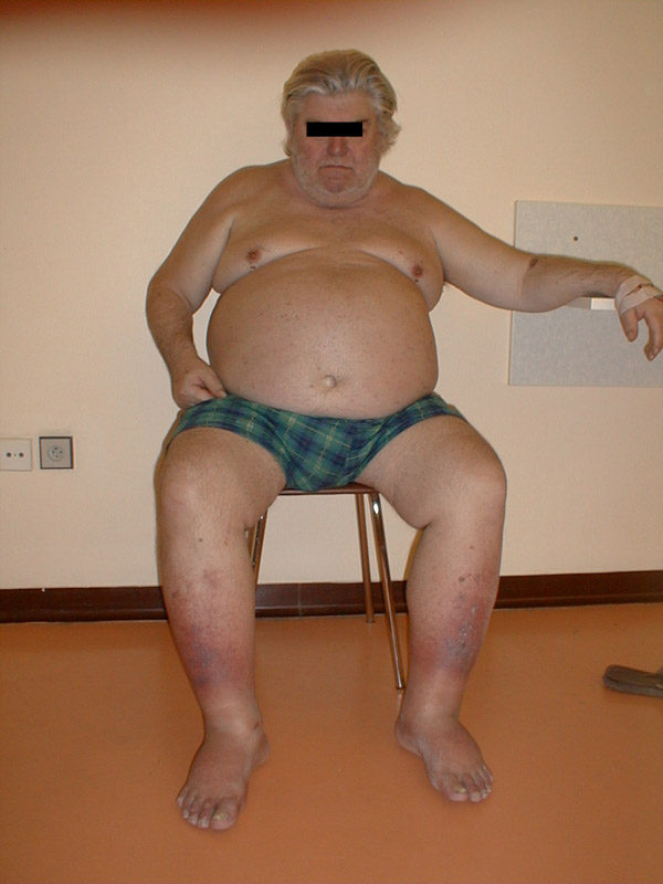
Symmetrical
cardiac oedemas
of lower
extremities
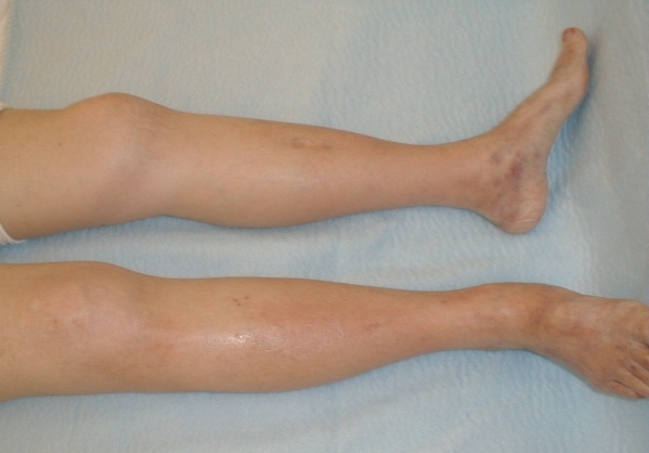
Wrinkled skin
of the lower
extremity after
oedema subsidence
Right-sided cardiac
decompression in
cor pulmonale
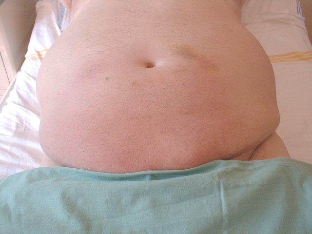
Anasarca
Nails
are generally strong, smooth, resistant and of distinctive appearance and colour.
Fragile and fraying nails are most common in thyrotoxicosis
and sideropenic anaemia.
Spoon-shape bent nails (koilonychia) occur in thyrotoxicosis.
Spherical nails accompany
congenital heart disorders, chronic pulmonary diseases; less frequently
can be found in hepatic cirrhosis as a part of clubbed fingers
(the shape of wrist watch glass).
"White" (hepatic) nails occur
in hepatic cirrhosis (the white part of the nail, so called
lunula occupies a significant part of the nail area).
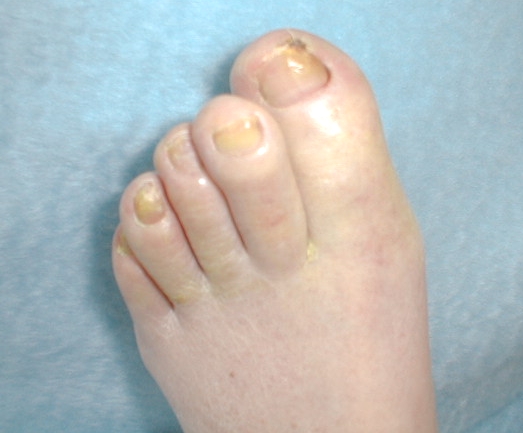
Nails deformed with uneven surface,
thick, changed in colour (particularly on toes) are affected by mycosis
(onychomycosis).
Your notes, observations, and proposals are welcome either via e-mail at the address
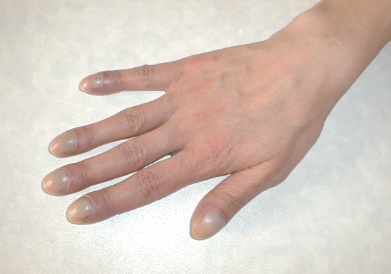
Detail -
clubbed fingers
with cyanosis
in central part in
congenital
heart disorder
Spherical nails