Examination of the heart
Observation
Percussion
Palpation
Auscultation
Auscultation findings in cardiac defects
Inspection, Percussion, Auscultation - is the most important,
Palpation. The other examination techniques are only supplementary.
Observation
 |
Shape of the thorax (kyphoscoliosis - cor pulmonale, kyphoscolioticum) |
| |
Postoperative scars |
| |
Visible beat of the apex (in left ventricle hypertrophy and dilatation, in hypertension, ischaemic heart disease, front wall aneurysm following a myocardial infarction, aortic defects, mitral insufficiency). |
| |
Systolic retraction of the intercostal space in adhesive pericarditis |
Extracardiac symptoms found by inspection:
Percussion
Provides a general orientation to determine the size of the heart (better X-ray picture of the heart + lungs, echocardiogram). Determination of the left heart margin - it should not exceed the midclavicular line.
Palpation
Under normal conditions, the beat of the heart apex is felt in the 4th - 5th intercostal space on the medial side of the midclavicular line.
Pathological findings:
| |
In left ventricle dilatation, the beat of the heart apex is shifted the left and downwards. |
| |
In left ventricle hypertrophy the apex beat is ascending. |
| |
In right ventricle dilatation and hypertrophy, the heart apex is shifted to the left, simultaneous occurrence of the systolic rising of the sternum and pulsation in the epigastrium (because the hypertrophic right ventricle leans against the front thoracic wall). |
| |
In extensive aneurysm or dyskinesia of left ventricle front wall, systolic pulsation can be felt along the left margin of the heart. |
Whirl is the palpation correlation of the murmurs. The following whirls can be palpated:
![]()

Kyphoscoliotic chest
Gibbus
![]()
Arcus senilis cornae
![]()
Xanthelasmata
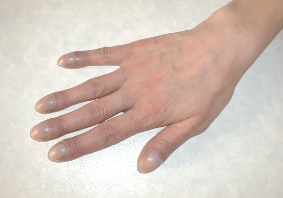
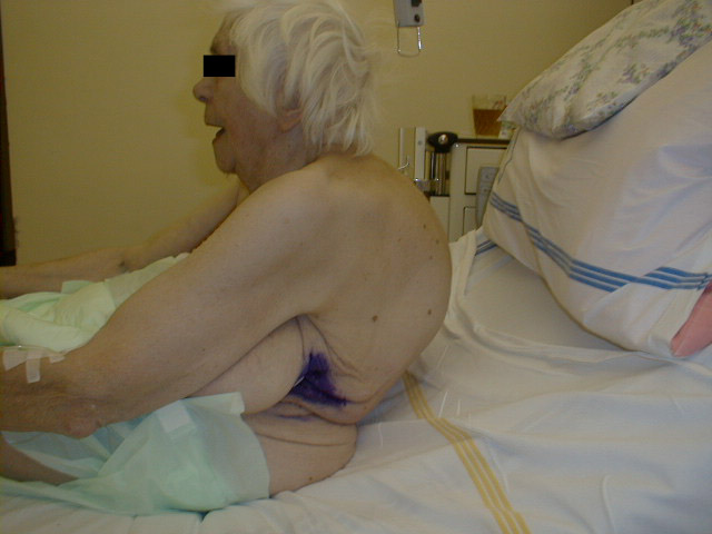
Digital clubbing
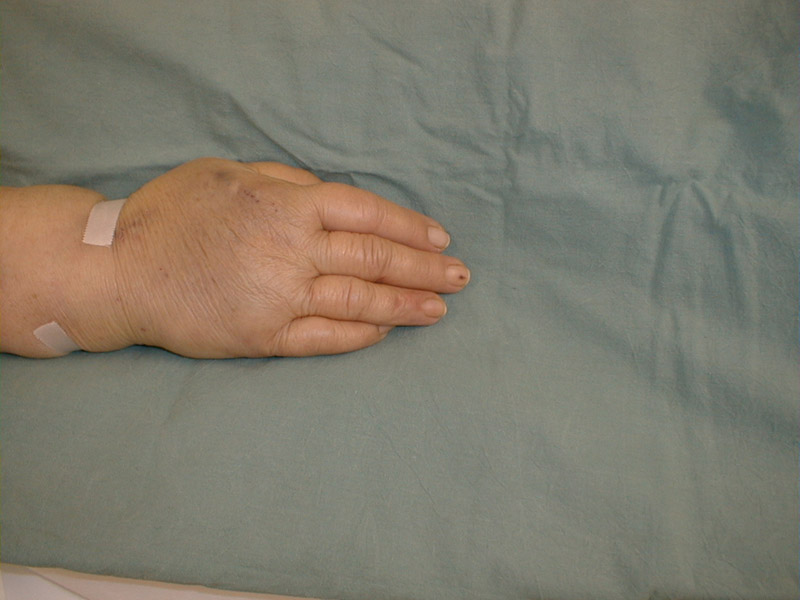
Splinter haematomas
Auscultation
Auscultation points on the chest
II right intercostal space close to the sternum
II left intercostal space close to the sternum
IV - V left intercostal space near the sternum
the intersection of IV and V intercostal space and the midclavicular line, the region of the heart apex
There are two types of stethoscopes
used - membranous (transmits better high frequency sounds and bell-shaped
(for listening to low frequency sounds and murmurs, e.g. the 3rd
sound, 4th sound, and diastolic mitral and tricuspid murmurs).
Normal cardiac sounds:
example
a); example
b)
Auscultation is of great significance - the most important physical
examination of the heart (see the picture Auscultation points
on the chest, sounds).
Ist sound
This is produced by closing of the mitral and then tricuspid valves
at the beginning of ventricular systole.
IInd sound
This is produced by closure of the semilunar valves, firstly the aortic
and secondly the pulmonary (changes with respiration). In expiration,
both components are getting closer together; in inspiration they are moving
away - the physiological segregation of the 2nd sound.
It is due to the fact, that in inspiration, the negative intrathoracic
pressure is intensified, the return to the right heart increases
and the increased stroke volume of the right ventricle prolongs
its ejection and therefore leads to a delayed closure of the pulmonary
valve - e.g. in BRTN (block of the right Tawara´s node) in ECG, in
atrioseptal defect etc.
NOTE - in pathological conditions, the paradox split of the 2nd
sound occurs if the left ventricle is overloaded and its systole
is prolonged (e.g. aortic stenosis, ischaemic heart disease, BLTN - block
of the left Tawara´s node in ECG) - closure of the aortic valve
is delayed, firstly the pulmonary, then the aortic component.
Therefore, in inspiration the 2nd sound is paradoxically
split, in inspiration the two components are moving away (i.e. the aortic
component and the pulmonary one physiologically), so that they merge.
IIIrd
sound
It is audible in patient lying on the left side, at the apex,
with the bell-shaped stethoscope.
It is produced by vibrations of the ventricular myocardium in the phase
of rapid filling of the ventricles at the beginning of diastole;
it is always produced, but as it is of low frequency character, it is
audible:
| |
In young patients in physiological conditions or |
| |
In elderly patients in pathological conditions it corresponds with low frequency -protodiastolic gallop (in failing heart). |
IVth sound
It is audible in the patient lying on the left side, at the apex, with the bell-shaped stethoscope.
It is elicited by vibrations of the ventricular myocardium on ejection of the blood into the ventricle during the ventricular systole at the end of diastole; but it is absent in atrial fibrillation!!!
It is found in young healthy people or in elderly patients as the presystolic gallop.
Concurrence of the 3rd and 4th sounds in cardiac insufficiency is called the summation gallop.
NOTE! IT IS ALWAYS AN IMPORTANT SIGN OF THE LEFT VENTRICULAR FAILURE!
Most frequent arrhythmia - - atrial fibrillation
Systolic clicks are accessory sounds produced during the systolic phase by the valvular activity.
| |
Ejection aortic click corresponds probably with sudden distension of the aortic valve. It occurs in early systole and is of a sharp clicking character. It is best audible in the apex region in dilatation of the aortic root, regardless of the cause of dilatation. |
| |
Systolic click may occur also during the midsystole or late systole, it sounds like a short, dry whip crack and can be multiple. It is caused by an abnormal function of the mitral valve and it is considered a sign of mitral valve prolapse. |
Ejection aortic and systolic clicks are often confused. Systolic click occurs later during the systole and in inspiration approaches the 1st sound.
Murmurs
Murmur - an acoustic phenomenon produced by vibrations of the valvular apparatus or another structure, if the laminar blood flow is replaced by turbulent circulation.
Classification of the murmurs based on localisation in the cardiac cycle:
| |
Systolic can
be functional (e.g. in childhood, in anaemia, in hyperkinetic circulation,
neurasthenia, thyreopathy, febrile conditions, stress) or of organic
aetiology. Murmurs are further classified according to a more
precise time specification into:
|
||||||||
| |
Diastolic
murmurs are always of organic aetiology (i.e. pathological!); they
are classified into:
|

Based on the character given by the prevailing frequency of vibrations, the murmurs are classified into:
| |
Harsh (rough) |
| |
Bellows (blowing) |
| |
Machinery |
| |
Musical (high-pitched character) |
| |
Continuous |
| |
Crescendo |
| |
Decrescendo |
| |
Crescendo - decrescendo. |
According to the intensity 6 murmur grades can be recognised:
| |
1st grade - hardly audible murmurs |
| |
2nd grade - murmurs soft, but audible |
| |
3rd grade - murmurs of middle audibility |
| |
4th grade - loud murmurs |
| |
5th grade - very loud murmurs, audible upon minimum contact of the stethoscope with the thoracic wall |
| |
6th grade - distant murmurs, i.e. audible without placing the stethoscope on the chest. |
4th to 6th grade murmurs are usually connected with the palpable whirl. The 2/6 murmurs indicate the 2nd grade intensity out of the given six grades.
Loudness of the murmur is usually proportional to the velocity of blood stream between the two cavities. Velocity of the blood movement depends on the pressure gradient over the ostium, on the shape of the ostium and size of the minute volume. In general, loud murmurs occur in a higher gradient, smaller ostium, or greater minute volume - e.g. a small ventricular septal defect elicits a very loud systolic murmur (great pressure gradient over the defect).
Auxiliary manoeuvres to improve murmur audibility:
| |
In exertion, murmurs are intensified, but in heart failure they remain unchanged. |
| |
Diastolic murmur in mitral stenosis is better audible in the position on the left side and following exercise. |
| |
Diastolic murmur in aortic regurgitation is examined in the sitting patient, slightly bent forwards. |
| |
Murmurs from the right heart are intensified in inspiration and they get reduced in Valsalva manoeuvre. |
| |
Left heart murmurs are accentuated in expiration. |
Accentuation of the sounds:
| |
Accentuation of the 2nd sound over the aorta is a manifestation of hypertension in the systemic circulation. |
| |
Accentuation of the 2nd sound over the pulmonary artery is a manifestation of hypertension in the pulmonary circulation |

| |
In pericardial exudation |
| |
In emphysema. |
Pericarditis
a) Dry pericarditis cracking sound like walking on newly fallen snow.
b) Exudative pericarditis
occurs in:
| |
acute myocardial infarction |
| |
viral infection |
| |
as a component of systemic disease |
| |
as postpericardiotomic syndrome (following cardiac surgeries). |
Your notes, observations, and proposals are welcome either via e-mail at the address