Picture 46

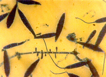
This picture shows Microsporum canis.
Microsporum canis
Morphology: Microconidia are long, clavate and borne along the hyphae. Macroconidia are large, fusiform with thick, rough walls. Colonies grown on Sabouraud's Dextrose Agar (SDA) show abundant, white aerial mycelium becoming buff in colour. A bright yellow orange pigment develops. Non-pigmented and glabrous variants occur.
Route of transmission: Direct contact
Investigations: KOH smear and culture from skin scrapings, discoloured hairs and kerototic debris
Diseases:
Infects, grows and remains confined to keratinous structures in the body.
1. Foot infection (Athlete's foot, tinea pedis), the hands are less commonly infected
2. Scalp infection (tinea capitis), can sometimes lead to an intense baggy suppuration known as a kerion
3. Nail infection (onychomycosis, tinea unguium) presents as white, discoloured nails or chalky, crumbling nails
Treatment:
Topical antifungals, eg. clotrimazole, miconazole, or ketoconazole for mild infections.
Systemic griseofulvin for more severe or unresponsive cases.
Oral ketoconazole for cases unresponsive to griseofulvin.