Examination of the abdomen
Observation - inspection
Percussion
Palpation
Examination of organs
Auscultation
Ascites
Examination per rectum
Methods of physical examination: observation
(inspection), percussion, palpation, and auscultation.
For orientation in the abdominal area topographic division
by lines is used:
 |
Horizontal - running below the costal arcs and connecting the flat parts of pelvic bones. |
| |
Vertical - running along the external margins of the straight abdominal muscles. |
The regions created are called:
Another possibility is to divide the abdomen into quadrants by means of vertical and horizontal lines running through the umbilicus into the right upper and lower quadrants, and left upper and lower quadrants.
The abdomen is examined in a recumbent patient with bent knees, in a quiet place. The examiner comes from the right, during the examination he/she should be sitting.
Observation (inspection)
is used to assess the level of the abdomen in to the thorax, symmetry, and progress of the breath wave.
Based on the nutritional condition, the physiological abdomen is the level or below the level of the chest. The navel is pulled in typical location. The breath wave proceeds bilaterally to the groin.
In addition to the abdomen, the inspection should be focused on the assessment of possible extra-abdominal disease manifestations in other locations.
General inspection

| |
Position
|
| |
Skin
|
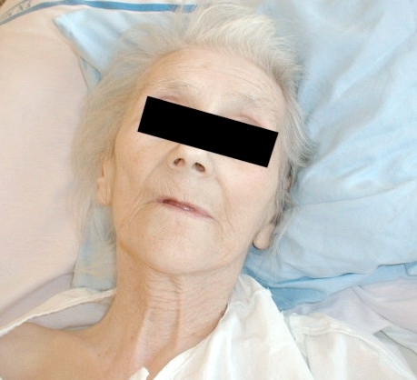
Icterus

Icterus
Icterus of the
sclerae and skin
of the face

Icterus

Spider nevi
in the face
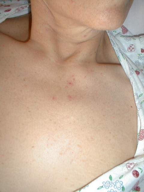
Spider nevi
Extra-abdominal inspection
Head
| |
Pale conjunctivae in anaemia. | ||||
| |
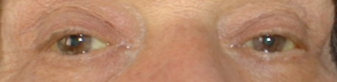
Yellow sclerae in icterus. | ||||
| |
Freckles surrounding eyes, mouth, and nose wings - occur in Peutz-Jeghers syndrome. | ||||
| |
Lips
|
Oral cavity
| |
Foetor ex ore - hepatic in liver failure (resembles the smell of mice). | ||||||
| |
Yellow-coloured palate in icterus. | ||||||
| |
Tongue
|
Lower extremities
| |
Hypoproteinaemic oedemas - perimalleolar or of a greater extent occur in liver cirrhosis, malabsorption syndromes etc. |
| |
Erythema nodosum is manifested on the crura in patients with idiopathic intestinal inflammations (idiopathic proctocolitis, Crohn's disease). |
Upper extremities
| |
Palmar erythema occurs in liver cirrhosis. |
| |
Dupuytren's contractures in palms are more frequent in patients with cirrhosis. |
Icterus of the
sclerae
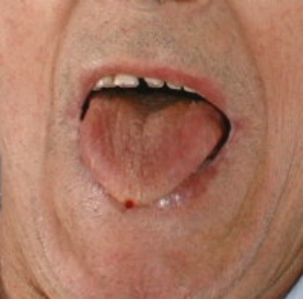
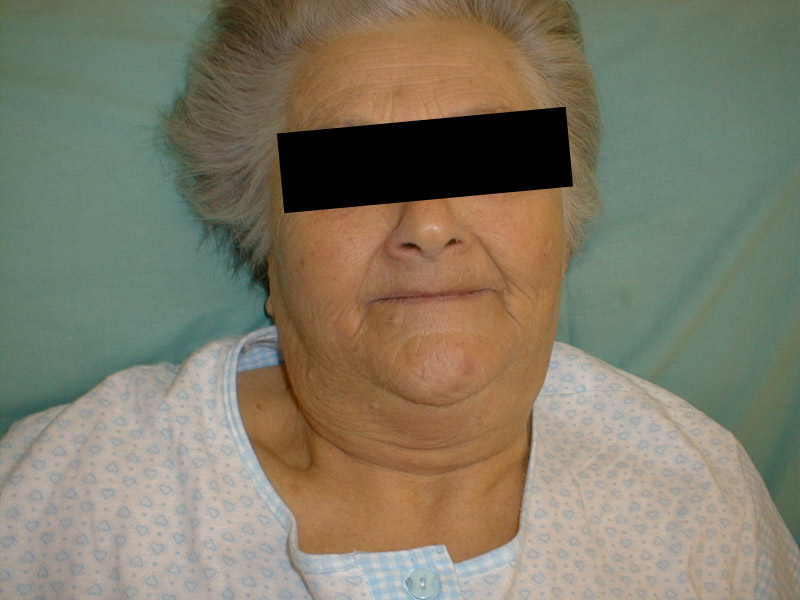
Dried-up tongue

Dried-up tongue
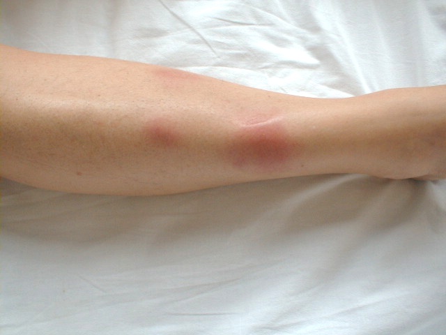
Erythema nodosum

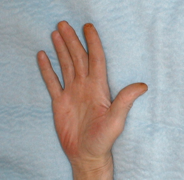
Dupuytren's
contracture,
palmar erythema,
tattoo of the
forearm + detail
| |
Navicular retraction occurs in extreme cachexia in tumours of the digestive tract. |
| |
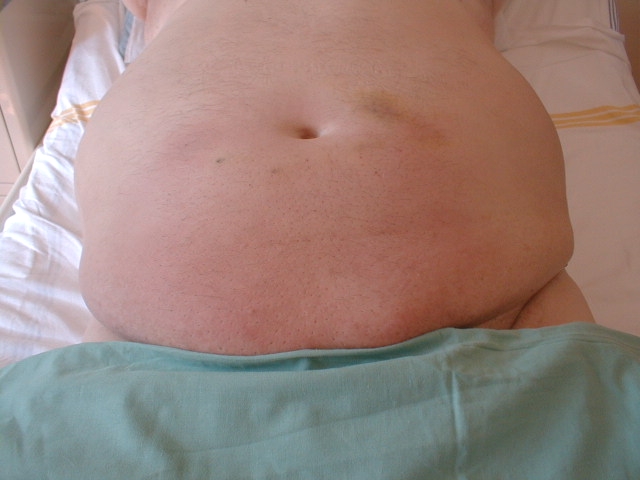
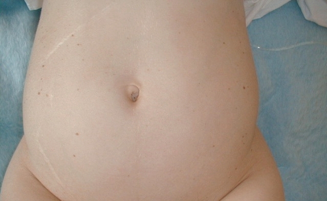
Above the level of the chest - it occurs in obesity, meteorism, pregnancy, and ascites, where abdominal shape is changed according to the patient’s position. |
| |
Breathing movements do not proceed through the abdominal wall in localised or diffuse peritonitis. |
| |
Visible pulsation of the abdominal aorta can be observed in thin patients or in aorta dilated by aneurysm. |
Colour of the skin
| |
Diffuse yellow in icterus, de-colouring is slower compared to the plasmatic level of bilirubin. |
| |
Paraumbilical violet (Cullen's sign) occurs due to propagation of retroperitoneal haematoma in severe acute pancreatitis. |
| |
Blue - haematomas of various age in haemorrhagic diathesis, related to subcutaneous application of heparin or insulin. |
| |
Pigmentation in the extent of linea alba in Addison's disease or after radiotherapy. |
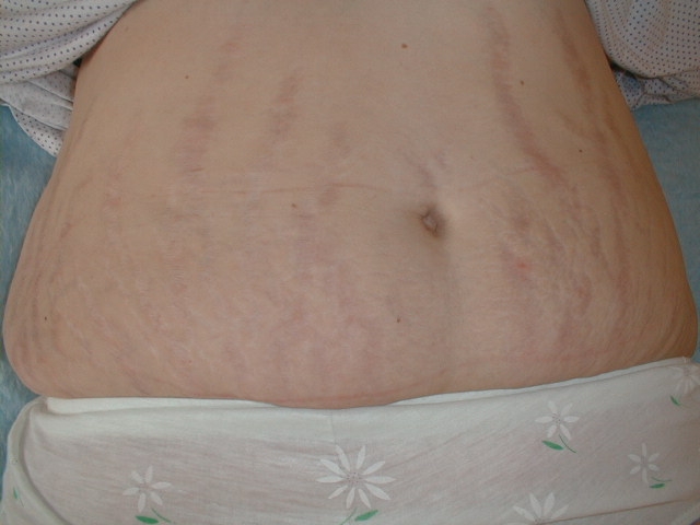
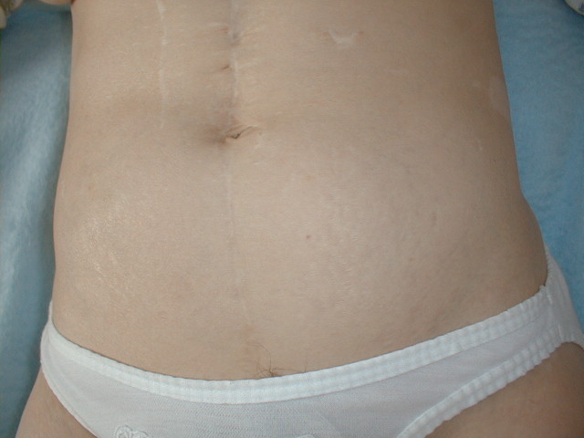
Striae
| |
Pearly striae are formed by the rapid distension of the abdominal wall in extension of the volume of the abdomen due to ascites, obesity, or pregnancy. |
| |
Violet in Cushing's syndrome. |
Venous pattern
"Caput medusae" - the veins radially converge to the navel or are visible in lateral parts of the abdomen. Both findings occur in portal hypertension.
Anasarca
Means advanced generalised effusion of the epidermis. The fluid is gathered also in the abdominal, thoracic, and pericardial cavities. It occurs in advanced right heart failure, hepatic cirrhosis, and serious hypoproteinaemia.
Postoperative scars
have typical localisation according to the type of operation. The most frequent are:
| |
After the upper middle laparotomy (surgeries of the stomach and duodenum, gallbladder, and biliary duct). |
| |
After lower middle laparotomy (gynaecologic, obstetric and urologic surgeries). |
| |
After the combined laparotomy (extensive abdominal surgery). |
| |
Right subcostal region (operation of the gallbladder). |
| |
In the right hypogastrium (appendectomy). |
| |
Suprapubic area (gynaecologic surgeries). |
| |
After the right-sided and left-sided lumbotomy (kidney surgery). |
| |
Combination of the mentioned scars with small scars of irregular shape (operations connected with drainage). |
| |
Short scars in various locations after diagnostic or therapeutic laparoscopy. |
The colour of the scar indicates its age (red-pink - recent surgery, skin-coloured scar - of older date). The complicated healing can result in formation of a hernia in the scar. In some patients, keloid scars can be found.
Physiological abdomen is symmetrical.
Pathological features that can be seen:
| |
Overall arch (bulge) in obese patients, in meteorism, iliac disorders, and in ascites (the shape of the abdomen changes relative to its position) |
| |
Local bulge due to cysts, hernias, diastases of the straight abdominal muscles, tumours, enlarged liver, or spleen, distended full stomach and/or intestine, and urinary bladder. |
| |
Hernias occur most often in the navel, groin, and postoperative scars (the size fluctuates depending on the intra-abdominal pressure). |
| |
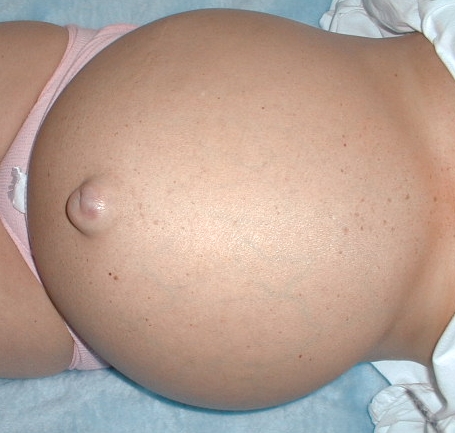
Eversion of the navel occurs in extensive ascites. |
| |
Peristalsis of the stomach and intestine is usually visible in pylorostenosis or intestinal obstruction (ileus). |
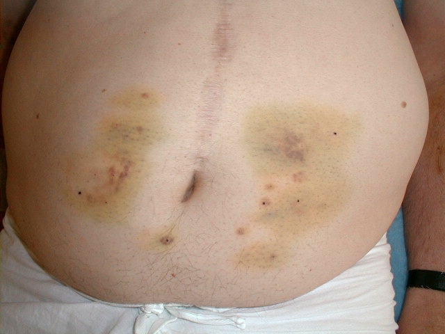
Abdomen - scar
following the upper
middle laparotomy
+ haematomas
after the s.c.
application of
low-molecular heparin
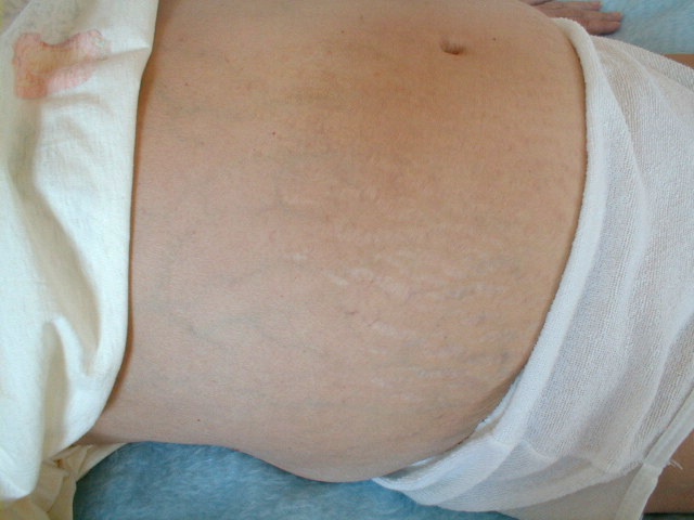
Pearly striae
Violet striae
Caput medusae
ascites,
eversion of the
navel, collateral
venous pattern
Anasarca -
effusion of the
abdominal wall
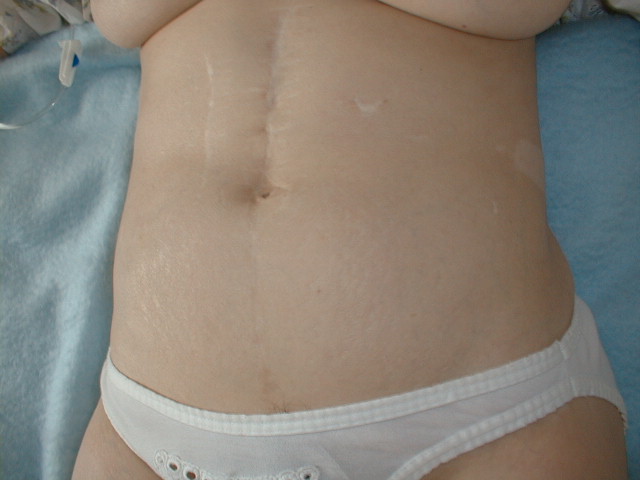
Scar after the
upper middle
laparotomy
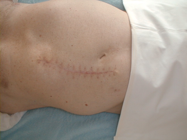
Scar after the
gallbladder
surgery
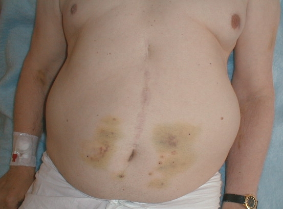
Abdomen - scar
after the upper
middle laparotomy
+ haematomas
after the s.c.
application of
low-molecular heparin
Abdomen - scar
after the upper
middle laparotomy,
vertical scar
along m. rectus
+ scars following
the drainage + striae
on the surface
of the abdomen
Scar following
appendectomy and
cholecystectomy
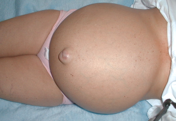
Overall arch
(bulge) of tje
abdomen, eversion
of the navel
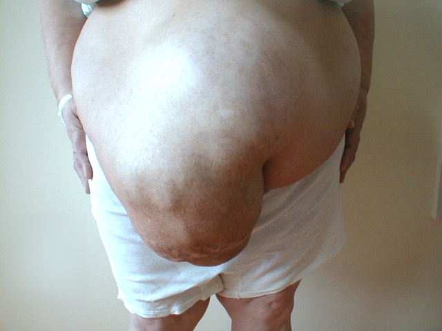
Hernia, obesity,
monstrous
ventral hernia
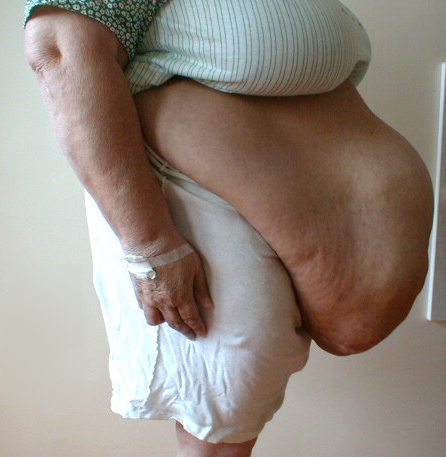
Obesity,
monstrous
ventral hernia,
and ascites
hepatic cirrhosis
![]()
Arch in the
epigastrium -
hepatocellular
carcinoma
with ascites