Observation - inspection
Percussion
Palpation
Examination of organs
Auscultation
Ascites
Examination per rectum
Liver
is not visible, its right lobe does not exceed costal arch, left lobe
reaches to one third to a half of the distance between xiphoid
process and navel (umbilicus), its movements correspond with breathing,
the edge is clearly defined, the surface is smooth, with soft
consistency, painless. The liver is wide 8 to 12 cm in the midclavicular
line (detectable by percussion).
The examination can be more difficult in obesity and distended colon.
Size:
Hepatomegaly - in cardiac decompensation, hepatic cirrhosis,
steatosis, or haematological diseases.
Edge
 |
Blunt, rounded – hepatic venostasis. |
| |
Sharp, thin - hepatic cirrhosis. |
| |
Uneven, rugged - metastases, hepatic cirrhosis. |
Surface
| |
Smooth - venostasis, hepatitis. |
| |
Uneven - macronodular cirrhosis, metastases. |
Consistency
| |
1st grade - soft, elastic - healthy liver. |
| |
2nd grade - tougher in venostasis, in inflammatory and infiltration liver diseases. |
| |
3rd grade - tough, inflexible – hepatic cirrhosis. |
| |
4th grade - very tough (rock) is found in cancer infiltration. |
Soreness originates in acute enlargement of the liver (hepatomegaly) due to tension of the capsule (acute venostasis, infectious hepatitis, acute cholangitis).
Pulsation of the liver is present in serious tricuspid valve insufficiency of the heart.
Hepatojugular reflux is produced by the manual pressure on the venostatic liver; it is manifested by the increased filling of cervical veins.
Riedel´s lobe is characterised by extension of the lateral part of the liver lobe downwards.
Gallbladder
is neither visible, nor palpable, nor painful.
Differential diagnostic problems, mainly those originating from the palpation, are caused mostly by Riedel´s lobe, liver metastases, infiltration of the omentum, colon cancer, and the right kidney ptosis.
Spleen
is neither visible nor palpable; during examination it shows associated movements (synkinesis) with breathing.
(The patient is examined lying on the right side, the physician stands behind the patient's back at the left bed side.)
Difficulties in examination are caused by obesity, tension of the abdominal wall, meteorism, ascites.
Local evaluation of the finding is – can be confounded with the enlarged left lobe of the liver, tumour of the left kidney and adrenal gland, tumour of the splenic (lienal) flexure or the cauda of the pancreas.
Splenomegaly can be visible in case of the extreme enlargement in asthenic patients.
Soreness
pronounced in perisplenitis joined with splenic infarction (mitral stenosis, infectious endocarditis), simultaneous frictional murmur can be palpable and audible.
Kidneys and urinary duct
are commonly neither visible nor palpable; they are not painful.
Diagnostic problems in physical examination are caused by difficult differentiation of hydronephrosis, polycystosis, and renal tumour, in relation to the colon or gallbladder tumour, and on the left side, to splenomegaly.
Stomach
in a healthy person it is not detectable either by inspection or palpation, it is not painful.
Pancreas
Due to its location in the retroperitoneum it is not accessible either by inspection or palpation.
Sigmoid colon
In a healthy man it is palpable in the left hypogastrium as a firm, smooth, cylindrical tube, sometimes tender on palpation.
| |
Tenderness - in inflammation (diverticulitis, idiopathic proctocolitis), or irritable colon. |
| |
Resistance, fixation to the base - tumour, an advanced process with the infiltration into the surrounding. |
Caecum
is sometimes accessible by means of deep palpation resulting in detection of a painless wider cylinder, slightly mobile against the base.
| |
Tenderness - corresponds with inflammations (idiopathic proctocolitis, Crohn's disease), or tumour. |
| |
Resistance - mostly due to tumour. |
| |
Distension - probably related to ileus condition. |
| |
Variable picture - with temporary finding of spastic contracted intestine - occurs in irritable colon. |
Appendix
requires extraordinary attention. In a healthy individual it is not painful even on deep palpation. It is palpable on the boundary line of the external and middle third of the umbilicospinal line (McBurney´s point).
Appendicitis is usually associated with the following findings:
Small intestine
is neither visible nor palpable. The jejunum is localised mostly in the left mesogastrium; the ileum in the right meso- and hypogastrium.
| |
Tenderness, rather diffuse - intestinal infection (enteritis), Crohn's disease, or distension. |
| |
"Stiffening of the loops" - caused by the intensive peristalsis in the place of obstruction (ileus). |
Auscultation
in routine examination of the abdomen is not usually used.
Intestinal phenomena, present in healthy people, are audible approx. 15x per minute. Loud sounds are sometimes of distant character.
The examination is performed in case of suspected disorder of the intestinal passage or vascular disorder (aortic aneurysm, arterial stenosis).
Peristalsis
Splashing sounds are heard in examination of the undulation above the distended organ, e.g. the stomach in pyloric obstruction or gastroparesis.
Friction murmurs are heard in perihepatitis and perisplenitis in dependence on breathing. They are detected rather by auscultation than palpation.
Vascular murmurs are heard above the stenotic sections of the abdominal aorta, renal arteries, lineal artery, and celiac plexus.
Auscultation is indicated in hypertension, in suspected abdominal aorta aneurysm.

Ascites
Ascites means the presence of free liquid in the peritoneal
cavity. It is detectable by physical examination if it exceeds 2000 ml.
The shape of the abdomen is changed in dependence on the body position.
Ascites is detectable by means of percussion. The limits are cranial-concave, variable in accordance with the patient's position. Major ascites is associated with the presence of undulation phenomenon (palpable rush of the liquid produced by the impact against the contralateral abdominal wall). The patient is examined both in recumbent (lying) and standing positions.
Ascites occurs in decompensated liver cirrhosis, advanced congestive heart failure, nephrotic syndrome, portal vein thrombosis, and carcinoma of the peritoneum. It is necessary, using differential diagnostics, to differentiate ascites from the encapsulated fluid in the abdominal cavity (ovarian and pancreatic cystis), which could be difficult or even impossible by means of a mere physical examination.
Per rectum examination
Is used as a routine preventive oncologic examination (prostate, rectal carcinoma), in developed or suspected enterorrhagia and melena and sudden abdominal emergencies.
The patient is in position "on all fours", supine, lying on the left or right side. Careful examination requires releasing of the abdominal wall.
Examination procedure
| Localisation | Physiological Findings | Pathological Findings |
| vicinity of the rectum | without changes | irritation of the skin, excoriation, fibromas, fistulas, fibrosed nodules, bluish transparent external haemorrhoids, tense thrombotic haemorrhoidal nodule, postoperative scars |
| anus | without changes | half-open (hypotonia of the sphincters) |
| distended anus | without changes | temporary haemorrhoids fissure, excoriation |
| indagation per rectum | unfeasible in anal fissure (intensive pain) | |
| tonus of the sphincters | adequate | increased: neurolability, fissure of the anus, the irritation of the canal, thrombosis decreased: atonia (at old age) |
| soreness | absent | present: fissure of the anus, irritation |
| anal canal | patent, free | small mobile formation - thrombotic internal haemorrhoid, polyp, anal papilla, tumour |
| rectal ampulla | spacious, the wall elastic, usual presence of the stools | infiltrated wall - with a tumour or inflammation (painful) |
| lumen of the intestine (within reach of approx. 8 – 10 cm) | without any pathological finding | polyp, tumour or lumen obstruction |
| cervix of uterus | smooth, painless, of medium tough consistency | condition after hysterectomy, tumour |
| prostate | symmetrical, soft, central line well palpable, painless | increased : - hypertrophy asymmetrical tough, uneven, the central line impalpable - carcinoma soft, tender - prostatitis |
Your notes, observations, and proposals are welcome either via e-mail at the address
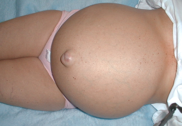
Caput medusae
ascites, eversion
of the navel,
colateral venous
pattern

Limits of ascites

Variable limits
in accordance
with patient's
position